Hemodialysis Access Care
What is HemoDialysis?
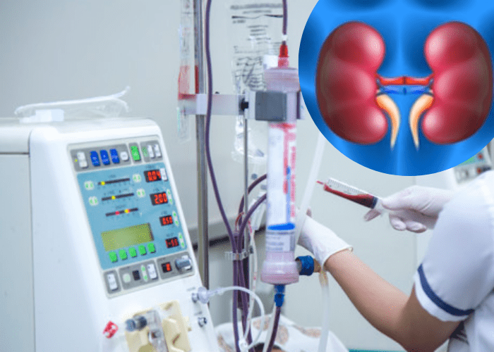
In medicine, dialysis is the process for patients who experience kidney failure. During such failure, the kidney fails to cleanse the blood of wastes, including urea or any extra fluid. Hemodialysis, also called dialysis, is used to withdraw blood from the body into a machine. This procedure uses a special membrane (Dialyzer) to filter wastes and extracts excess fluid from the blood. It also restores the electrolyte balance in the blood. Dialysis usually takes about three hours and occurs thrice a week. Avail the best Hemodialysis Access Care in Mumbai with us.
What is Hemodialysis Access?
Hemodialysis Access - also called "Vascular Access" - is a way to withdraw blood for hemodialysis. Due to the need to withdraw faulty blood from the body and replace it, a means for accessing the patient's blood circulation through hemodialysis becomes necessary. There are three different types of procedures for this, some of which are used interchangeably: Dialysis Fistula, Graft and Catheter. All of these techniques can remove and supply large amounts of blood into the body at the same time--about one quart per minute.
What is a Fistula?
Fistula is a connection established between blood vessels (artery & vein). It is considered the most desirable form of hemodialysis vascular access. Surgeons create an arteriovenous fistula & connect an artery to a vein and make the blood flow smoothly in the upper arm or forearm.
With time, typically one to three months, the vein expands and gets ready to receive the needles used to withdraw and supply blood during dialysis. A fistula can last for years if it matures well, i.e. if it develops well.. During the time that a fistula is growing, if hemodialysis is necessary, another form of vascular access will be required, usually through a catheter.
What if the fistula does not develop?
A non-maturing or non-developing fistula happens in up to one-fourth of patients. There are two reasons for a non-maturing fistula: either narrowing of a vein or too many competing veins. Interventional radiologists can both open up the narrowed vein with a balloon (Balloon Angioplasty) or close off the competing veins using several methods. About three-quarters of people with non-maturing fistula will profit from one or both of these procedures and have their fistula develop.
What is a Dialysis Graft?
In some patients, the arteries or veins are not suitable for making a fistula and for such patients, a graft (or shunt) can be applied as an alternative form of Dialysis Access. This alternative is basically a plastic tube that connects an artery to a vein. Unlike fistulas, the grafts do not need to "develop" and are ready for use in most cases by four weeks after the placement. During this waiting period, a catheter may be necessary for dialysis. The drawbacks of grafts are that they do not last nearly as long as the fistulas do and can develop narrowing and clotting more often. Besides, there is something with grafts, something which does not happen very often with fistulas -- they can get infected.
By making appropriate choices in terms of screening techniques, narrowing veins with grafts can be detected before they clot. These involve self-examination, check-ups by the interventional radiologist and measuring flows during dialysis. Once the medics discover an abnormality, you need to be scheduled to have it treated by interventional radiology as promptly as possible. Patients must keep their appointments with interventional radiology in time to avoid clotting. The patient needs not worry in case of clotting as it can be handled efficiently by an interventional radiologist.
What is a Hemodialysis Catheter?
The catheters used for hemodialysis are small tubes that are placed under the skin. They are considered the least desirable form of dialysis access. Catheters come in two ways: a short-term (non-tunneled) and longer-term (tunneled) form. Its best use is to provide limited access to dialysis for patients whose kidney function is expected to improve. It is also used on patients whose kidney function is not likely to improve but who have a graft or fistula in place as they are awaiting it to develop.
An interventional radiologist inserts a catheter through one of the large veins, usually the jugular, into larger veins that are located in the center of the chest near the heart. This procedure lasts less than an hour and can be done as an outpatient. The best outcomes with catheter placement are achieved through the use of imaging guidance, including ultrasound, to inject a needle into the vein and X-rays to lead the accurate placement of the catheter.
They hold the advantage of being able to be used for dialysis instantly after they are placed. Patients are also fond of them because the needle sticks are not necessary to remove and replace blood during dialysis, which is common with graft or fistula procedures. However, catheters come with their set of significant disadvantages and challenges, including:
-
There are complications including the risk of infection and blockage.
-
One of the higher rates of complications of catheters involve central venous stenosis wherein the veins they are placed into can get clot off or develop constrictions. In fact, with particular chest veins called the subclavian veins (just under the collarbone), the risk of clotting or narrowing is approximately 50%. Therefore, subclavian veins should never be used for catheters except in exceptional cases when all other veins have been used up. Patients can help to prevent this complexity by not allowing their doctors to use the subclavian veins for dialysis catheters, instead insisting on the jugular veins where this complication is pretty uncommon (less than 10%).
-
Most of the patients find catheters uncomfortable and/or deformed.
Despite the issues with catheters, patients may require them for a short period as a substitute by the time a fistula develops or a graft heals. Ordinarily, it should take less than three months for a fistula and a month for the graft.
Besides implanting dialysis catheters, interventional radiologists also treat difficulties with catheters like infection and clotting. These problems are generally treated by replacing the catheter for a new one in a brief outpatient method lasting less than an hour.
What are the screening tests for failing dialysis access?
Screening for failing dialysis access can be done in several ways, out of which self-examination is the simplest. The best and easiest way is USG doppler, which can be done by our experts. If there is poor flow detected, then a prompt visit to our experts for a fistulogram and balloon angioplasty as needed.
What if problems are detected with my graft or fistula?
While a fistula is regarded as the most suitable kind of access, some complications can occur, which include, vein narrowing, stenosis, and clotting or thrombosis. An interventional radiologist, like our experts, can treat both of these problems with remarkable consequences. Operating the vein while it is narrowed but not clotted provides the best results and takes the least amount of time.
What if my dialysis access is clotted?
When the graft or fistula is clotted, interventional radiologists use a variety of methods to dissolve or remove the clots. First, X-ray pictures (Fistulograms) are taken, which show the areas of stenosis. Then a procedure called an "angioplasty," or fistuloplasty, a balloon is inserted to clear the obstructed area(s) in the vein. A clot can either be removed with drugs that dissolve it or with mechanical devices that remove it or break it into tiny pieces.
These procedures are all done using conscious sedation and local anaesthesia (numbing medicine). After angioplasty, blood flow usually returns to normal. The balloon angioplasty may need to be repeated periodically, usually every six months. When angioplasty fails, interventional radiologists have other options available to use. A small metal tube called a "stent" can be inserted in the same procedure as the angioplasty depending upon the location of the narrowing. This process is used quite uncommonly and more often in the chest than the arms. In the case of angioplasty failure, the patient may be referred to a surgeon for further procedures that include "revision of the graft or fistula".
